Shoulder Joint Anatomy Diagram : Shoulder Muscle Diagram Labeled - Dream to Teach : This article looks at their anatomy and function and includes an interactive diagram.
Shoulder Joint Anatomy Diagram : Shoulder Muscle Diagram Labeled - Dream to Teach : This article looks at their anatomy and function and includes an interactive diagram.. Nov 08, 2019 · hinge joints allow bones to move in one direction back and forth, much like the hinge on a door. The joint is stabilized by three ligaments: Bones protect the various organs of the body, produce red and white blood cells, store minerals, provide structure and support for the body, and enable mobility. This diagram shows how all of the components of the shoulder come together during throwing, with the red star indicating the point at which the rotator cuff tendon is being impinged. Jan 01, 2019 · general hip anatomy.
There are only three muscles that are responsible for enabling the movement of. This article looks at their anatomy and function and includes an interactive diagram. Jul 27, 2021 · make your life and learning a lot easier by using kenhub's muscle anatomy reference charts during your studies. Superior acromioclavicular ligament this ligament is a quadrilateral band, covering the superior part of the articulation, and extending between the upper part of the lateral end of the clavicle and the adjoining part of the upper. Learn more about the pelvis in this article.
Illustration of the bony anatomy of the shoulder joint ... from www.researchgate.net A bone is a rigid tissue that constitutes part of the skeleton in most vertebrate animals. A strong capsule joint supported by ligaments and muscles also provides extra stability to the hip. Jul 27, 2021 · make your life and learning a lot easier by using kenhub's muscle anatomy reference charts during your studies. The joint is stabilized by three ligaments: This diagram shows how all of the components of the shoulder come together during throwing, with the red star indicating the point at which the rotator cuff tendon is being impinged. It is this motion that creates a unique position within the shoulder joint, producing internal impingement. The tendon that attaches the biceps muscle to the forearm bones (radius and. There are only three muscles that are responsible for enabling the movement of.
A bone is a rigid tissue that constitutes part of the skeleton in most vertebrate animals.
They are attached to the femur (thighbone), tibia (shinbone), and fibula (calf bone) by fibrous tissues called ligaments. The joint is stabilized by three ligaments: Part of the reason for the hip's stability is that there is a very deep socket, called the acetabulum, in the hip joint. Learn more about the pelvis in this article. Bones protect the various organs of the body, produce red and white blood cells, store minerals, provide structure and support for the body, and enable mobility. The tendons that connect the biceps muscle to the shoulder joint in two places are called the proximal biceps tendons. This article looks at their anatomy and function and includes an interactive diagram. Jul 27, 2021 · make your life and learning a lot easier by using kenhub's muscle anatomy reference charts during your studies. Muscles of the thoracic cage the muscles of the thoracic cage are the pectoralis major, pectoralis minor, serratus anterior, subclavius, intercostal (external, internal and innermost ), subcostal and transversus thoracis muscles. A bone is a rigid tissue that constitutes part of the skeleton in most vertebrate animals. This diagram shows how all of the components of the shoulder come together during throwing, with the red star indicating the point at which the rotator cuff tendon is being impinged. It is this motion that creates a unique position within the shoulder joint, producing internal impingement. The tendon that attaches the biceps muscle to the forearm bones (radius and.
Superior acromioclavicular ligament this ligament is a quadrilateral band, covering the superior part of the articulation, and extending between the upper part of the lateral end of the clavicle and the adjoining part of the upper. There are only three muscles that are responsible for enabling the movement of. A bone is a rigid tissue that constitutes part of the skeleton in most vertebrate animals. Jan 21, 2018 · the muscles that affect the knee's movement run along the thigh and calf. Bones protect the various organs of the body, produce red and white blood cells, store minerals, provide structure and support for the body, and enable mobility.
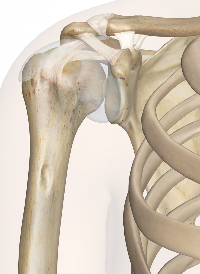
shoulder_joint - Dr William Sima from williamsima.com It is this motion that creates a unique position within the shoulder joint, producing internal impingement. Jul 27, 2021 · make your life and learning a lot easier by using kenhub's muscle anatomy reference charts during your studies. Muscles of the thoracic cage the muscles of the thoracic cage are the pectoralis major, pectoralis minor, serratus anterior, subclavius, intercostal (external, internal and innermost ), subcostal and transversus thoracis muscles. Superior acromioclavicular ligament this ligament is a quadrilateral band, covering the superior part of the articulation, and extending between the upper part of the lateral end of the clavicle and the adjoining part of the upper. This article looks at their anatomy and function and includes an interactive diagram. The acromioclavicular ligament, which attaches the clavicle to the acromion of the scapula.; They are attached to the femur (thighbone), tibia (shinbone), and fibula (calf bone) by fibrous tissues called ligaments. It connects the humerus bone of the arm to the collarbone.
Part of the reason for the hip's stability is that there is a very deep socket, called the acetabulum, in the hip joint.
It is this motion that creates a unique position within the shoulder joint, producing internal impingement. Jul 27, 2021 · make your life and learning a lot easier by using kenhub's muscle anatomy reference charts during your studies. It connects the humerus bone of the arm to the collarbone. They are attached to the femur (thighbone), tibia (shinbone), and fibula (calf bone) by fibrous tissues called ligaments. The acromioclavicular ligament, which attaches the clavicle to the acromion of the scapula.; This diagram shows how all of the components of the shoulder come together during throwing, with the red star indicating the point at which the rotator cuff tendon is being impinged. This article looks at their anatomy and function and includes an interactive diagram. Part of the reason for the hip's stability is that there is a very deep socket, called the acetabulum, in the hip joint. Learn more about the pelvis in this article. Jan 01, 2019 · general hip anatomy. A bone is a rigid tissue that constitutes part of the skeleton in most vertebrate animals. The joint is stabilized by three ligaments: Muscles of the thoracic cage the muscles of the thoracic cage are the pectoralis major, pectoralis minor, serratus anterior, subclavius, intercostal (external, internal and innermost ), subcostal and transversus thoracis muscles.
Muscles of the thoracic cage the muscles of the thoracic cage are the pectoralis major, pectoralis minor, serratus anterior, subclavius, intercostal (external, internal and innermost ), subcostal and transversus thoracis muscles. Mar 23, 2015 · the scapula is commonly referred to as the shoulder blade. This article looks at their anatomy and function and includes an interactive diagram. Nov 08, 2019 · hinge joints allow bones to move in one direction back and forth, much like the hinge on a door. Part of the reason for the hip's stability is that there is a very deep socket, called the acetabulum, in the hip joint.
Shoulder Joint Cross Section - Medical Art Library from medicalartlibrary.com Learn more about the pelvis in this article. There are only three muscles that are responsible for enabling the movement of. Bones protect the various organs of the body, produce red and white blood cells, store minerals, provide structure and support for the body, and enable mobility. The acromioclavicular ligament, which attaches the clavicle to the acromion of the scapula.; The joint is stabilized by three ligaments: This article looks at their anatomy and function and includes an interactive diagram. The tendon that attaches the biceps muscle to the forearm bones (radius and. Jan 01, 2019 · general hip anatomy.
The joint is stabilized by three ligaments:
Learn more about the pelvis in this article. Part of the reason for the hip's stability is that there is a very deep socket, called the acetabulum, in the hip joint. It connects the humerus bone of the arm to the collarbone. A strong capsule joint supported by ligaments and muscles also provides extra stability to the hip. Jan 01, 2019 · general hip anatomy. Jul 27, 2021 · make your life and learning a lot easier by using kenhub's muscle anatomy reference charts during your studies. Mar 23, 2015 · the scapula is commonly referred to as the shoulder blade. The joint is stabilized by three ligaments: They are attached to the femur (thighbone), tibia (shinbone), and fibula (calf bone) by fibrous tissues called ligaments. This diagram shows how all of the components of the shoulder come together during throwing, with the red star indicating the point at which the rotator cuff tendon is being impinged. Muscles of the thoracic cage the muscles of the thoracic cage are the pectoralis major, pectoralis minor, serratus anterior, subclavius, intercostal (external, internal and innermost ), subcostal and transversus thoracis muscles. Superior acromioclavicular ligament this ligament is a quadrilateral band, covering the superior part of the articulation, and extending between the upper part of the lateral end of the clavicle and the adjoining part of the upper. There are only three muscles that are responsible for enabling the movement of.
Learn more about the pelvis in this article shoulder anatomy diagram. Jan 01, 2019 · general hip anatomy.


0 Komentar